You’ve probably used a microscope before: usually, the sort of low-power microscope that pervades grade schools and simple collegiate science labs put together just to fulfill the most basic of requirements to graduate. But these days, high-end microscopy is changing science, allowing researchers to see and study things never-before seen in new and fascinating ways.
The microscopes used in cutting-edge research are massive: often filling rooms or buildings, and costing tens or hundreds of millions of dollars to purchase and keep in perfect working condition. Some require vacuum to work properly; others require super-chilled temperatures requiring liquid nitrogen and ethane. While NASA spends millions on super-specialized rockets and stations to explore space, other scientists, many government-funded, use specialized microscopes to turn the eye of science in the other direction: to the smallest particles already on earth.
These microscopes are used in dozens of research disciplines, from physics to microbiology to chemistry, studying the how and why of proteins, cells, and atoms.
Structural Microscopy
Cryo EM (or cryo-electron microscopy) is a relatively new discipline in microscopy, which focuses on looking at biological samples in their native environment. A long-standing problem of microscopes which look at biological samples in the past has been that samples would need to be manipulated, such as stained, divested of their natural surroundings and are therefore less-scientifically relevant. With cryo EM, samples are literally cryogenically frozen and then imaged in their native state, meaning that scientists can study the actual structure of sub-cellular proteins, organelles, and other items.
For example, a scientist studying how a certain bacteria infects and causes disease can use cryo EM to study and capture the natural structure of the proteins which harm human cells, or the bacterial structures which translocate those proteins. The fact that cryo EM ensures these biologicals remain as in-tact as possible means that scientists can hasten the process from study to cure, and dramatically improve the accuracy of their results. But that’s not all!
Crytomography is a discipline within cryo EM which allows scientists to take thousands of pictures of what they’re studying, to create full 3D renderings. Before the advent of cryo EM, often getting complex 3D renderings would require long years of research trying to crystallize the proteins and structures being studied… when it was possible to do so at all! Cromotomography can cut that time in half, and allow scientists to render samples without fixative or alteration.
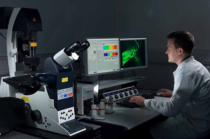
Taking Images of the Never-Before Seen
There’s a burgeoning trend of using high-resolution microscopy images as art. But what does it take to get these images?
Super resolution confocal microscopy is the new standard, which allows scientists to see images at a resolution higher than the diffraction limit. Roughly ten high-profile microscopy techniques, including PALM and STORM all use this method of microscopy, which can be employed to follow fluorescent particles and microorganisms, and allow scientists to study into the nanodimension.
Often, the computer interface of large microscopes will be outfitted to take images. However, this can be a complex proceeding, and not all microscope computers will afford their users a GUI. In other cases, microscopes will be outfitted with ancillary options like the DinoLite Microscope Eyepiece which acts as a camera independent of the microscope.
Atomic Force Microscopy
Some scientists want to study things even smaller than the sub-cellular level. For this, they’ll use atomic force microscopy, which allows them to view particles more than 1000 times better than the optical diffraction limit. That means you’re seeing the literal atoms which make up the sample.
This is a stunning achievement of science, which allows scientists to study the atomic behaviors of matter under many different conditions. This sort of research is useful in the development of nanotechnology, especially where it relates to biomedical applications, such as new methods of drug delivery.