The brain, that intricate network of almost one hundred billion neurons and the same number of non-neuronal cells – astrocytes and oligodendrocytes, among others – has captivated and challenged the scientific community for centuries.
To understand how neural circuits allow us to get excited by the smell of a perfume, feel empathy or display complex behaviors such as creativity or ethical decision-making, we must first understand the structure and function of the different types of brain cells and their relationship.
Of course, as you can imagine, identifying so many billions of brain cells is a scientific and technological challenge, even more so if we want to characterize each type of cell in particular. Well, this monumental effort has just been carried out successfully.
Finally a census of brain cells
In order to make such an ambitious project a reality, the brain cell census research network, called BICCN , was created in 2017. This network includes more than thirty laboratories from various disciplines. Its goal is to identify, characterize and map each type of brain cell in humans, non-human primates and mice.
The BICCN studies, thanks to the use of the most advanced technologies that until now were only applied to animal models, have borne their first fruits: they have revealed the detailed cellular composition of the human brain, both in adulthood and during its development. The findings are detailed in a series of twenty-four scientific articles , mostly published in the prestigious journals Science and Science advances .
And they have done it at three different levels of study: transcriptional , which tells us the function of cells through the expression of their genes; the epigenetic , which reveals how these genes are activated or deactivated by age and environmental factors; and the functional level , which refers, for example, to whether neurons excite or inhibit other neurons.
The integration of these results shows that, as expected, in addition to the variation between brain regions, there is also variation between the brains of each person. That is, there is not a single prototype of the human brain, but rather a wide genetic range and response to the environment, both in healthy individuals and in different disease states.
Two atlases and two comparative analyzes
The enormous and complex research at the level of analysis of individual cells has provided very interesting results. Firstly, two atlases have been generated: one of individual cells from the adult human brain, and another of individual cells from adult non-human primates ( macaques and marmosets ).
Likewise, two comparative analyzes have been presented, one of individual cells between human and non-human primate brains, and another between individual cells during brain development in both humans and non-human primates .
Finally, the function and distribution of human neuronal cell types and their comparison with those of mice have been analyzed and modeled . As they would say in my country: “Almost nothing.”
Detailed description of more than three million cells
Among the most important results is the detailed description of more than three million individual brain cells (including more than two million neurons) from almost one hundred different areas of the adult human brain.
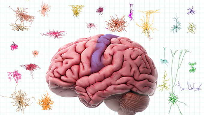
The findings indicate that the brain is not homogeneous at all. Although all brain cells share the same DNA, each of them uses different genes in different amounts. That gives rise to an absolutely astonishing level of cellular diversity and specialization . Each brain area contains a specific set of cell types and in different functional states. In addition to helping us understand how the brain works, knowing them will be of great clinical use in relation to diseases whose brain alteration may be different depending on the person who suffers from it, such as brain tumors, epilepsy or multiple sclerosis, to name a few.
Interestingly, the most unique neurons are found in the primary visual cortex (V1), which has become the epicenter of fascinating discoveries about how we interpret and perceive the visual world around us. This brain region is not only an image processor, but also an impressive mosaic of cells that together create the rich tapestry of our visual experience, allowing us to discern shapes, colors and movements with astonishing precision.
This work lays the foundation for understanding how variations in cellular structure can influence our ability to process information and perform various cognitive functions.
Small but significant differences from our close relatives
Furthermore, other research also reveals surprises in human brain cells when we compare them with those of our closest relatives, chimpanzees and gorillas. Although we share a basic brain cell structure, our neurons use different genes to connect and form circuits in the brain. This detail indicates that small changes in neural connections could evolutionarily boost our cognitive abilities, such as complex reasoning and the creation of advanced languages.
Adding to these results, the team revealed an interesting neural similarity between chimpanzees and gorillas, despite the fact that chimpanzees and humans share a more immediate ancestor. This highlights the exceptional nature of the brain biology that makes us human, unfolding a range of possibilities such as the invention of tools, the composition of majestic symphonies and the perception of delicate sensitivity in poetry.
Implications in developmental disorders such as autism
In relation to the study of the development of the human cerebral cortex, which unfolds through our prenatal stage and continues for many years after birth, more than 700,000 cells from 169 tissue samples from 106 donors have been thoroughly analyzed .
Thus, it has been possible to establish how various cells develop and differentiate in the brain, including the neurons that are responsible for emitting electrical signals, those that regulate them, the glial cells that are the “caretakers” of the neuronal environment, and the that make up our cerebral blood vessels, all of them being fundamental pieces in the majestic puzzle of our brain machinery.
Regarding the implications that these findings have on developmental disorders such as autism, this work presents us with a perspective on how small changes in this complex dance of cellular development can lead to conditions that profoundly affect social and communicative interaction. For example, by understanding more about how neurons and glial cells develop and communicate with each other, we can begin to unravel the mysteries of why, in some people, this process differs and how this can impact the way we who perceive and interact with the world.
As if that were not enough, the study illuminates the subtle but significant differences in gene expression between girls and boys with respect to autism, providing a prism through which to examine why this disorder shows different rates of incidence and manifestation between genders.
Regardless of the enormous value of each of the published results, the interdisciplinary effort demonstrated here allows progress towards the common goal of knowing the development and functioning of the brain that makes us human . In addition to opening the doors to a new era of research into the origin of neurological diseases.
Author Bio: Francisco José Esteban Ruiz is a Full Professor of Cellular Biology at the University of Jaén