Human Anatomy is a basic subject in all studies in health sciences. Knowledge of the human body is key to developing good clinical practice: it improves understanding of pathology and skill in certain procedures in different disciplines, such as medicine, nursing, physiotherapy or psychology, among others.
Until a few years ago, their learning was based on dissecting corpses and viewing images in books. Today, virtual dissection has revolutionized the way we learn it.
Brief history of learning anatomy
The word “anatomy” comes from Greek and means to cut again (“ana” means to return, and “tomos” to cut), referring to its main learning method, dissection. Anatomy was already a science in Ancient Greece. One of its notable characters was Herophilus, the first doctor to dissect corpses , considered the father of anatomy.
Later, as the greatest representative of Ancient Rome in this area, Galen appears , who stated that a doctor without anatomical knowledge is like an architect without plans.
After a few years in the Islamic Golden Age , authors such as Muhammad Al-Razi stood out in the field of neuroanatomy, Ibn Al-Haytham in optics and Ibn al-Nafis, who explained pulmonary circulation, paving the way for William Harvey for many centuries. after.
After the Middle Ages, historical works began to appear in text form such as De Humani Corpis fabrica by Andrés Vesalius or paintings such as The Anatomy Lesson of Doctor Nicoales Tulp by Rembrandt . From here on, all that social interest in this matter began to be collected and reflected.
Around 1800 the demand for corpses grew, leading to the controversial practice of illegal exhumation of bodies. In fact, in the United Kingdom it was regulated with the Anatomy Act of 1832 .
Innovations in the 20th and 21st centuries
During the late 19th and 20th centuries, technological innovations such as x-rays, magnetic resonance imaging and CT scans appeared that allowed us to visualize the inside of the human body.
Finally, due to the advancement of all 3D visualization and modeling technologies, virtual anatomy visualization systems appear in the 21st century. They are tools that make a virtual copy of a human body or a real digitized corpse. The covid-19 pandemic was a turning point for its implementation in many universities that did not have this technology.
In fact, there was talk of the possible end of the use of cadavers for learning anatomy, due to the possible risks of contagion of covid-19 .
What is virtual dissection?
Virtual dissection allows human and animal anatomy to be taught using advanced digital technologies. It differs from traditional physical dissection in that cadavers or specimens are used.
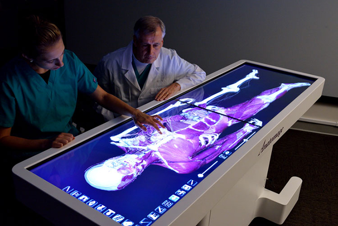
Among these tools is Anatomage , a virtual dissection tool that has appeared in scientific research since 2018 , and is already being used in some Spanish universities.
Interaction with these virtual dissection tables is similar to a digital tablet, but on a large scale. Which means that the tactile experience is lost (it is similar to using a mobile application), but you can execute actions that we could do with a corpse, such as making cuts, or visualizing a real structure in all planes of space.
This type of dissection has five notable characteristics:
- Visualization: Allows a detailed 3D view of anatomical structures. You can view layered tissue, organs and systems from multiple views and angles.
- Interactivity: You can “cut”, rotate, explore different parts of the body with a click. Facilitates active learning and experimentation.
- Accessibility: No laboratory or corpses needed. It can be accessed from anywhere, so it requires few resources and can be learned remotely.
- Realism: despite being a simulation, these tools offer greater realism every day, using real anatomical data to increase their precision.
- Personalization: you can learn based on needs, personalizing learning. The content can be modified, made easier and more difficult depending on the student’s level.
Advantages of using virtual dissection
There are a number of advantages to these virtual dissection models:
- Safety and ethics: eliminates the need to use animal corpses or specimens. Therefore, possible negative situations that may arise from its manipulation are eliminated.
- Less emotional impact: Working with corpses can have a powerful emotional impact. Also eliminate the smell.
- Repetition and consistency: procedures and image views can be repeated as many times as desired. This is not possible in all traditional dissection practices.
- Cost-effectiveness: it is a more economical alternative in the long term because it is not necessary to renew or maintain bodies or laboratories.
- Constant evolution: with the rise of 3D models and artificial intelligence we are at the beginning of the development of these technologies.
Its drawbacks
Not everything was going to be advantages, among its disadvantages are:
- Lack of tactile experience: this type of experience, which is so necessary in other procedures, cannot be replicated.
- Dependence on technology: requires access to technology, which may not be fully democratized and technical problems may arise.
- Loss of realism: Traditional anatomists argue that direct experience with cadavers provides a deeper understanding of the variability and complexity of the human body.
Do you learn more with virtual dissection?
It has been observed that the attitude of students is more favorable when learning anatomy with real cadavers than with digital dissection tools. However, virtual dissection has proven to be effective as a complement to traditional anatomy learning.
Combining these two types of dissection can enhance learning and improve results . It has also been observed that digital tools help to visualize content in a more understandable and clinically applicable way.
Virtual dissection offers a detailed, three-dimensional view, reducing emotional and ethical impact and being cost-efficient. Although it experiences challenges such as lack of tactile experience and technological dependency, it represents a significant advance in anatomy pedagogy.
Author Bio: Daniel Sanjuán Sánchez is a Physiotherapist and Research teaching staff at the Faculty of Health Sciences at Universidad San Jorge, Associate Professor at the Faculty of Nursing and Physiotherapy at the University of Lleida. Member of the iPhysio research group at San Jorge University