Imagine someone who feels a lump on their side. They go to the doctor, distressed, and the doctor, upon palpating, says, “That’s not a lump, it’s a floating rib.” It’s a funny anecdote to tell at the dinner table (and joke about the person’s possible hypochondria) because it was easy for the specialist to identify the problem and reassure the patient. However, this wisdom, which we take for granted, has been acquired over time. After all, we expect that the doctors who diagnose, treat, and operate on us have acquired a deep understanding of the human body and its diseases.
But accessing that learning hasn’t always been easy. Before the internet, video, and even color photography, how was that experience formed? In a sense… with art.
Sculptors to train doctors
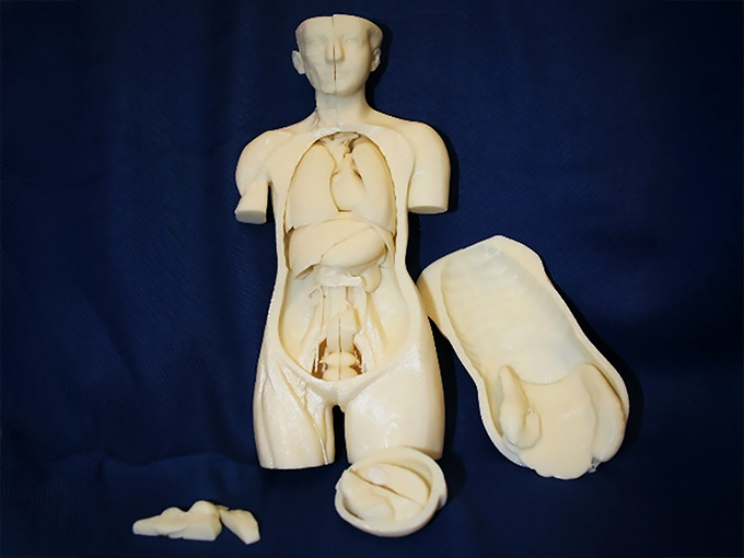
Along with dissections, anatomical models played a fundamental role in the teaching of medicine: they reproduced with great fidelity – in three dimensions, in color and at natural scale – the human body and its pathologies.
During the 18th and 19th centuries, it was common for medical institutions to assemble anatomical collections for educational museums. In Spain, this practice was formalized by a Royal Decree of 1862, which required the seven universities—eight from 1876 onwards—to create public positions for anatomical sculptors and their assistants, responsible for producing these pieces.
However, despite being the only sculptors with a permanent position at the university for more than a century, their work was undervalued and remains almost forgotten today.
The anatomical museums established in 18th-century Spain, linked to the royal colleges of surgery, were inspired by Italian models such as La Specola in Florence, with its famous wax figures by Clemente Susini. These pieces emphasized the anatomy of an often idealized body and, in the case of the anatomical Venuses , possessed an erotic dimension aimed at the male viewer.
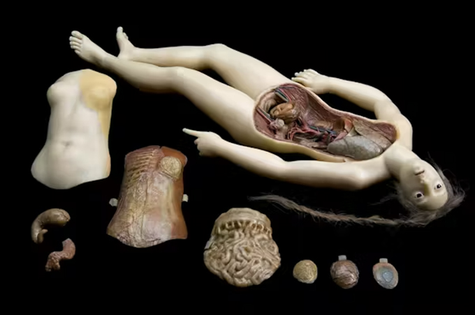
Anatomical Venus made by Clemente Susini, wax, 1780-1782. Museo di Palazzo Poggi, Bologna/Wikimedia Commons , CC BY
With the strengthening of medical schools and their museums, this approach shifted slightly. By the mid-19th century, the binary concepts of subjectivity and objectivity had solidified , with the latter becoming the ideal of medical practice; realism became its principal ally. In this context, anatomical sculpture focused on the direct representation of disease, encompassing everything from the most common to the most exceptional pathologies. Simultaneously, wax was gradually replaced by plaster, which was more economical and durable.
Professionals between art and medicine
Access to a position as an anatomical sculptor required dual training, both artistic and anatomical, and joint examination boards comprised of Fine Arts and Medicine experts. It was a difficult profile to maintain in a poorly recognized and underpaid post. As Dr. Ángel Pulido lamented in 1883, those who held the position “renounce being painter, sculptor, and doctor, to become a hybrid being, a mixture of all three at once,” lacking the prestige of any of the disciplines.
Even so, the precariousness of the art world led many sculptors to work in medical schools. Many of them achieved prominence in the art world, winning awards at national fine arts exhibitions, having their work acquired by the state, and being featured in museums such as the Prado and the National Art Museum of Catalonia, in addition to receiving commissions for public monuments. Their names appear in university archives and, occasionally, in the signatures of the surviving pieces.
Thus, the archives of the Complutense University of Madrid reveal the presence at the Madrid faculty of renowned sculptors such as Maximino Sala , Miguel de la Cruz , José Ortells , and José Pérez (“Peresejo”) . These are joined by artists linked to Santiago de Compostela, such as Juan Sanmartín and Francisco Asorey , and to Granada, with Francisco Morales . The most prominent case is Barcelona, where Juan Samsó , Rosendo Nobas , Torquato Tasso , Dionisio Renart , and Enric Monjo worked .
However, this anatomical facet barely appears in their biographies, as it is considered marginal in relation to “true” art. This is the case of Samsó, active at the Faculty of Barcelona between 1863 and 1878, who combined the production of anatomical models –tumors, malformations, and births– with successful participation in national exhibitions.
Nobas, his successor, created the Quadriga of Aurora , a large public monument bathed in gold, while also producing series in painted plaster depicting the before, during, and after of surgical operations. He also left a unique testament to the intersection of art and medicine in the marble tomb of the anatomist Jaime Farreras Framis. On it, a skeleton wrapped in a shroud appears with breathtaking realism.
Forgotten heritage
A quick glance at the inventories of 19th-century anatomical museums reveals that most of the specimens have been lost, either misplaced, deteriorated, or destroyed. Although the surviving collections have generally remained in the hands of universities, most of these museums have been dismantled. Among them all, the Javier Puerta Museum of Anatomy stands out as an exceptional case of survival.
In Barcelona, the preserved pieces ended up in the Museum of the History of Medicine of Catalonia , where they remain almost inaccessible to the public. The imminent closure of the Olavide Museum of dermatological models highlights the fragility of this heritage.
However, it is an essential legacy for understanding how the body and illness were conceived in the 19th century. And also, and no less importantly, for completing the history of sculpture in Spain.
Author Bio: Chloe Sharpe is Professor and Researcher in Art History, UDIT – University of Design at the Innovation and Technology